Saturday, April 26, 2008

Where the Wild Microbes Are: A New Theory on How Pathogens Survive Food Processing

ResearchBlogging.orgProduce borne diseases have recently been gracing the front pages of our media. Our , our and , our , and our and . Not to mention the countless tons of .

Common sense says that washing and proper handling of our food should simply be enough to prevent illness outbreaks. It has now been hypothesized that many bacteria were able to "hide" within and among the plant cells, protected by their sturdy cell wall. Or even that some pathogenic bacteria were able to enter the cells and remain protected from traditional washing methods.

An article in this month's Applied and Environmental Microbiology looks at a much different method of bacterial survival on produce. They hypothesize that these bacteria are taking refuge in various protozoa, and subsequently are protected from washing and other sanitation methods due to being held either within the cell or an exogenous cell-derived vesicle.

The authors used protozoal isolates from store-bought spinach and romaine lettuce. They measured the concentrations of various microbes from the surface, and cultured a set of naturally occurring protists from these samples.

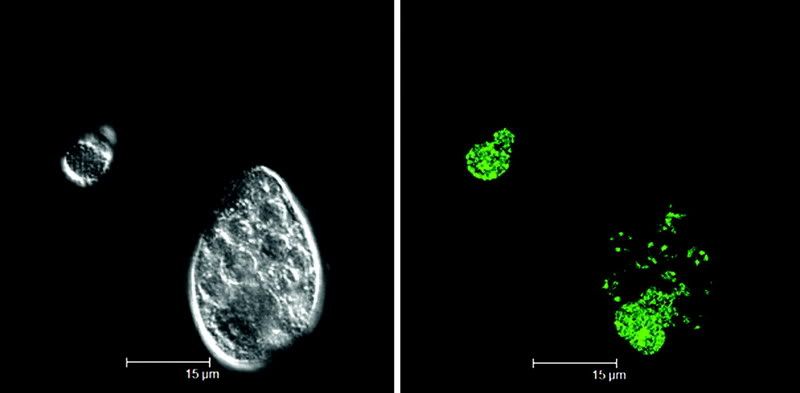
They show that not only are certain pathogenic bacteria (like ) able to be taken up by protozoa, but that in many cases, they survive to be ejected into the world around, surrounded by a vesicle. The flourescent image at above shows this event happening with E. coli 0157:H7 and a strain of Tetrahymena.
Previous studies have shown that the , even in harsh conditions.

This paper shows us that microbial ecology is much more complex than we perceive, and that simple and obvious solutions may not always be the best. We now know that wild protozoa living on our produce can sequester pathogenic bacteria, and furthermore wet produce allows vesicles to be created containing viable bacteria.

This makes me wonder about the current trend in grocery stores to spray there fresh produce with water at specific time intervals. If protists are doing what this study shows they are doing, and vesicles form on wet leaf surfaces, this practice may be something we must reconsider for our food safety.


Gourabathini, P., Brandl, M.T., Redding, K.S., Gunderson, J.H., Berk, S.G. (2008). Interactions between Food-Borne Pathogens and Protozoa Isolated from Lettuce and Spinach. Applied and Environmental Microbiology, 74(8), 2518-2525. DOI: 10.1128/AEM.02709-07

Happy Birthday Elio Schaechter!

would like to take a moment to wish Elio Schaechter at a very happy 80th Birthday.

Small Things Considered is a joint collaboration between Elio Schaechter and Merry Youle. These fine writers post on the wonders within the world of microbes. Theirblog is always entertaining and informative, and serves as a unique looking glass at "the Small Things."

Thursday, April 24, 2008

Oncolytics: Not just for Myxoma Virus


In a previous article, I discussed the use of against malignant gliomas.

Recently, I came across a post from , pointing to an article describing a as treatment against human sarcomas.

This continues to show us that viruses can be highly beneficial to our society. Antibacterial therapy and cancer therapy are just two of the many advances that viruses have been crucial in development.

It is my opinion that we should begin seriously exploring the potential therapies that Nature has already created for us, rather than concentrate on de novo synthesis of treatments. There are an infinite number of mechanisms and pathways in Nature that could provide groundbreaking technologies.

It is just up to us to find them and utilize them properly.

Tuesday, April 15, 2008

The Definition of Life; and, Taxonomy as We Know It

ResearchBlogging.orgIn order to make sense of our surroundings, we, as humans, constantly group things into discrete catagories--states of matter, times of day, types of clothes, etc.

Since , we have consistantly grouped life in boxes-within-boxes, based on physical characteristics. The advent of nucleic acid sequencing changed some of these early notions of taxonomy. But now, as we discover more about life, specifically of our microbial counterparts and their viruses, we are coming to the realization that Linnaein taxonomy is not sufficient. Especially if our motivation for using taxonomy is to deduce evolutionary relationships.

Viral taxonomy has some very specific shortfalls. Researchers in this , lay out these shortcomings. One striking point is that with viruses, structure alone is not sufficient to characterize a phage into species. The figure here shows two similar looking phages with very different DNA sequence. This concept could easily be applied to a variety of other viruses.

Another problem, is that species is defined as organisms that share a common gene pool. Viruses are constantly moving genetic information into and out of hosts. Many viruses have the ability to cross-infect more than one host as well. This means that the gene pool available to an organism crosses most borders of what we traditionally define as species.

The muddle within genetic taxonomy is getting a recent look. The discovery of MIcrobe MImic Virus () is a startling look at how complex this tangled web is.

A recent article in Nature Microbiology, discusses not only the question of taxonomy, but challanges our definition of life. Their definition presupposes that viruses are more complex than merely "parasitic nucelic acids." They propose that the presence of either a capsid or ribosomes be the primary classification system for life and that genomic contents completely define an organism.

The proposal consists of defining capsid-encoding organisms (the viruses), ribosome-encoding organisms (eukarya, prokarya, and archea), and "orphan replicons" (plasmids and ).

Whether or not this classification scheme will take root remains to be seen. There will certainly be critics, however, I think that the evidence available strongly supports the classification of viruses as a segment of life.

Raoult, D., Forterre, P. (2008). Redefining viruses: lessons from Mimivirus. Nature Reviews Microbiology, 6(4), 315-319. DOI: 10.1038/nrmicro1858

Further Reading Possibilities
Liaisons of Life: From Hornworts to Hippos--How the Unassuming Microbe has Driven Evolution
Microcosmos: Four Billion Years of Microbial Evolution


Monday, April 14, 2008

Seminar Time

You know you've sat through lectures at seminars or conferences that sounded like this.


Sunday, April 13, 2008

Monthly Book List, First Edition

Here are a variety of books that may interest some of you. Many of these are personal favorites, some are entertaining and educational, others just educational, but rife with knowledge.

The Hot Zone: A Terrifying True Story
Follow the story of the Ebola virus. This book brought me into the world of virology. Exciting and educational...not many people know that we had an Ebola virus outbreak right in Virginia.

The Demon in the Freezer: A True Story
Another book by Richard Preston, this time centering around the bioterrorist threat of smallpox and anthrax. Although quite embellished, this is an entertaining read and definatly worthy of its place on my bookshelf.

Biohazard: The Chilling True Story of the Largest Covert Biological Weapons Program in the World--Told from Inside by the Man Who Ran It
This book scared me when I first read it. Written by an ex-Soviet scientist who defected to the US, he tells us his story developing biological weapons of mass destruction. A great read, but not for the faint of heart.

The Coming Plague: Newly Emerging Diseases in a World Out of Balance
Another look at emerging infectious diseases. Although "The Hot Zone" started my interest in looking at how viruses work. "The Coming Plague" initiated my studies in where infectious diseases come from.


Molecular and Cell Biology Carnival #1

Announcement:
The inaugural Molecular and Cell Biology Carnival has been published. Hosted over at The Skeptical Alchemist, this edition highlights some current topics in MCB.

This is the first of (hopefully) many, so feel free to submit your articles for future inclusion over at Blog Carnival.

Enjoy!

Friday, April 11, 2008

A Fatty Acid Synthetase is Necessary for Active TB Infection

ResearchBlogging.org is the most common infectious agent in the world. , and a drastic number of these are increasingly antibiotic resistant. and are becoming more commonplace everyday, especially in regions that are combating HIV infections.

As such, it is incredibly important to study how this bug works. It is one of the most difficult organisms to work with due to a wide variety of factors. These include: slow growth rate, pathogenicity, very waxy cell walls, high levels of illegitimate recombination, and a bunch of other nuances that make the mycobacteria unique among its relatives.

One unique feature of the , is their unique staining pattern. It has been shown that when treated with (the most prescribed anti-TB drug), mycobacteria lose this pattern of staining. Therefore, one can extrapolate that their outer cell wall (filled with extremely long-chain lipids, called ) plays an important role in both the lifestyle of M. tb. and its pathogenesis.

Two genes have been identified as having roles in mycolic acid synthesis, kasA and kasB. KasA has previously been shown to be essential (J Bacteriol. 2005 Nov;187(22):7596-606.), with kasB serving a non-essential accessory function. This paper, from Bill Jacobs’ lab at AECOM, shows that although kasB is nonessential, it is necessary for acid-fastness AND it has a large role in pathogenesis.

They first knocked out kasB using a . Knockouts of kasB had a severe deficiency in colony growth and a large change in colony morphology.

Also, kasB mutants lacked acid fastness and were exceptionally more sensitive to .

Further biochemical analysis showed that kasB is likely involved in the production of specific mycolic acids.

However, the story gets more exciting. Immunocompetant mice that were infected with the kasB mutant, showed no signs of infection—there was no formation of granulomas and no mortality. In contrast, mice infected with wild-type died within 356 days. What is interesting to note is that although mice infected with kasB mutants did not show pathology, they still had measurable amounts of bacterial growth—indicating a persistent infection that was not able to become active (Fig. below).

The same study shows that kasB mutants act nearly identically to wild-type in mice that are immunocompromised. This means that mycolic acid production by kasB plays a role in the immune systems ability to hold M. tb. infections in check.

The authors suggest a variety of implications that their very exciting findings bring to light. For one, due to the phenotype, kasB mutants could be used specifically to study persistant infection. Secondly, kasB is clearly a candidate for deletion in an attenuated vaccine strain. Finally, and I think most importantly, this mutant shows a potential drug target for secondary drug therapy. Drugs that act on kasB or kasA could be developed in conjunction with rifampicin and isoniazid treatment.

XDR-TB cases are present in 41 countries, including the US. It is vital that we develop new lines of drugs and continue identifying drug targets. TB presents an event where scientists, public health departments, and drug industry must come together and work as a single entity. The amount of TB cases is ever increasing and I fear that the days of the sanitarium may once return to our country.


Bhatt, A., Fujiwara, N., Bhatt, K., Gurcha, S.S., Kremer, L., Chen, B., Chan, J., Porcelli, S.A., Kobayashi, K., Besra, G.S., Jacobs, W.R. (2007). Deletion of kasB in Mycobacterium tuberculosis causes loss of acid-fastness and subclinical latent tuberculosis in immunocompetent mice. Proceedings of the National Academy of Sciences, 104(12), 5157-5162. DOI: 10.1073/pnas.0608654104

Suggested Readings
Molecular Genetics Mycobacteria
Mycobacteria Protocols (Methods in Molecular Biology) (Methods in Molecular Biology)
The White Plague: Tuberculosis, Man, and Society
Timebomb : The Global Epidemic of Multi-Drug Resistant Tuberculosis


Thursday, April 10, 2008

Wild Bacteria That Eat Our Antibiotics? Of Course!

ResearchBlogging.org were invented by bacteria and fungi during thier conception in this universe. Used to control microbial niche environments, it wasn't until 1928 that (and subsequently and ) began the widespread use of the antibiotic to control bacterial infections in humans. And so, the antibiotic revolution began.

Subsequent use (and misuse) of antibiotics has given rise to various resistant strains. These are becoming a vast problem in the treatment of diseases that once were "easily" curable, including the well-publisized and , as well as many others.

This paper, coming out of Harvard University, describes the isolation of bacterial strains that can live on antibiotics as their sole carbon source. Current thought states that resistance in a bacterial population occurs because of exposure in clinical settings (or a popular theme of ). However, the authors show that even secluded environmental isolates have the ability to subsist on both natural and synthetic antibiotics.

Our antibiotics are just variations on themes we have seen in nature, and because of this, the environment represents a vast source of antibiotic resistance mechanisms that we have yet to discover. In niches exposed to antibiotics (ie. clinical pathogens) complete resistance is preferential. However, antibiotic metabolism (as described in this paper) would confer an advantage, even if it is only small in comparison.

I find it fascinating that in 11 different soils (urban, farm, and pristine), bacteria were present that could thrive on 18 different antibiotics as carbon sources! These bugs were not just living with the antibiotics, but were actually eating them!

We have much more to discover about antibiotic resistance mechanisms, this study clearly shows a direction that we can head to begin this task. Just as we looked to nature to develop antibioitcs, so must we look to nature to study how resistance works.


Dantas, G., Sommer, M.O., Oluwasegun, R.D., Church, G.M. (2008). Bacteria Subsisting on Antibiotics. Science, 320(5872), 100-103. DOI: 10.1126/science.1155157


Further Readings
Timebomb : The Global Epidemic of Multi-Drug Resistant Tuberculosis
Good Germs, Bad Germs: Health and Survival in a Bacterial World
Teaming with Microbes: A Gardener's Guide to the Soil Food Web


Monday, April 7, 2008

Phages With Horns?! What's Next?

ResearchBlogging.orgIn the world of phage, uniqueness rules. The total number of phages in the biosphere is dramatic, with estimates numbering the population at 1031! Despite such high numbers, since the discovery of phage no identical phage has been found in the environment twice. They have often been termed "Nature's Most Successful Experiment." Their genomes are in constant flux amoung themselves and their hosts. It therefore should no longer surprise us when we discover a phage that doesn't quite fit the mold.

In this paper by Pope, et al. they describe a phage (named Syn5) of the that comes equipped with a "horn." Described as a "slender elongated fibrous protrusion," the horn clocks in at 50nm in length. To put perspective on this, the horn is almost as long as the capsid is wide (60nm) and is longer than the tail (25nm). It is however, quite thin 10nm wide at its thickest point and 2nm at its thinnest.

The figure below is a cryoEM taken of Syn5, showing the fibrous horn structure directly opposite the tail.
Every Syn5 capsid has this protrusion, and it only occurs once in its specific location.

The purpose is yet unknown, however there is a running hypothesis. These phages live out in the open ocean where there hosts are not around in very high numbers. Therefore, it would be highly beneficial for the phage to increase its chances of finding and attaching to a host. The phage would not want to be oriented in the wrong direction as a host cell passed overhead. If this horn functions as a host recognition site, clearly it would increase the phage's chances to find a host cell.

To further amaze you, the vast majority of cyanophage

I can only imagine what we will find next as we continue to explore this small world. Truly,


Suggested Reading

The Bacteriophages

The Cyanobacteria: Molecular Biology, Genomics and Evolution


POPE, W., WEIGELE, P., CHANG, J., PEDULLA, M., FORD, M., HOUTZ, J., JIANG, W., CHIU, W., HATFULL, G., HENDRIX, R. (2007). Genome Sequence, Structural Proteins, and Capsid Organization of the Cyanophage Syn5: A “Horned” Bacteriophage of Marine Synechococcus. Journal of Molecular Biology, 368(4), 966-981. DOI: 10.1016/j.jmb.2007.02.046


Saturday, April 5, 2008

I Got You Phage

Back in August of 2006, I wrote and performed this song for a lab meeting to summarize my work I had done that summer and the previous year (All regarding a bacteriophage called BPs).

Some of the lyrics are hard to catch, you can find them below the embed. Enjoy!


Lyrics
Will I find you? Well, I don't know.
I won't find out until my will grow.
I scraped the dirt right off my shoe
You were there and , I found you.

(Chorus)
Phage, I got you phage. I got you phage. I got you phage.

They say you won't kill , you'll only infect
I can tell from your lack of
You all the bacteria we got

(Chorus)

You have in your
You have on your
And when your cruising round the town
when you're around.

So let them say your tail's too long
I don't care, I wrote the song
So put your little head by mine
It has the we can find

(Chorus)

from dirty sludge
I found you while eating fudge
I got of you
I still got work to do
I want you to , I'll watch it when you replicate
I found you, I won't let go
I found you, I love you so

Phage
I got you phage
I got you phage I got you phage I got you phage
~End Lyrics~


There will hopefully be some more microbiology songs coming soon (as soon as I get a camera up and running). With titles such as:
1. It's Gonna Be There (The E. coli song)
2. Brillant Dance of the Starvation Response
3. Phage in the Soil


Friday, April 4, 2008

Recombineering: A Practical Application of Phage Biology

ResearchBlogging.orgThe most obvious use of phages is of course direct phage therapy. Although exciting, there are many other uses of phage that are just as revolutionary, but tend to slip by. In my last article, I hinted at (Short for homology-dependent, recombination-mediated, genetic engineering) This is a method whereby phage proteins are used to catalyze homologous recombination. The ramifications are huge. Mutations can be made; whole genes and operons can be knocked out with relative ease, and much more. In fact, some may say that this recently described process revolutionized genetics.

Since this system was so successful in E. coli, the Hatfull Lab at the University of Pittsburgh set out to develop this system in the . Before this, genetic manipulation (especially gene knockouts) was highly difficult. M. tuberculosis is notoriously recalcitrant to genetic manipulation. This is due to a variety of factors including: its horribly slow growth rate, high pathogenicity, and its relatively high levels of illegitimate recombination compared to homologous recombination.

This paper, by vanKessel and Hatfull was published relatively recently (Jan 2007) and describes what is a great advance in mycobacterial genetics.

The first step was to identify potential recombination proteins encoded by phages. These proteins are presumed to be necessary for proper facilitation of lysogenic integration and DNA replication. Looking at the functions of the “Red” recombination system, (used for recombineering) one can find functional homologs in other systems. The Red system uses three proteins: exo, beta, and gam. Exo acts as a 5’-3’ dsDNA dependant exonuclease, beta acts to bind ssDNA and promote annealing, and gam functions to inactivate an alternative host recombination pathway (the RecBCD system).

Using this as the basis for comparison, one first comes across the Rac of E. coli. This prophage encodes two proteins called RecE and RecT that function identically to the exo and beta genes respectivly in the Red system, without significant sequence similarity. One then can look at the published mycobacteriophage genomes (30 at the time of publication, soon to be 50) and search for functional analogs of either the Red or Rac system. The authors found that in one phage of the 30, recET homologs were found. This phage, Che9c, contains two genes that are each around 30% identical to the Rac prophage RecE and RecT (at Che9c gp60 and 61).

Biochemical analysis showed that RecE and RecT function nearly identically to the Lambda phage exo and beta.

The authors placed Che9c gp60 and 61 onto a plasmid under an acetamide promoter (high expression of these genes under a constitutive promoter was determined to be toxic) and transformed into M. smegmatis (a fast growing, non-pathogenic relative to M. tuberculosis)

These cells could then be transformed with a dsDNA leuD knockout substrate containing 1,000bp homology on each side (500bp flanking sequence and 500bp leuD sequence) of a Hyg resistance cassette. Upon induction of the RecET homologs, allelic exchange was shown to occur at a measureable frequence (8x10^-4 recombinants per ug / cell competancy) by detecting the presence of leucine auxotrophs with hygromycin resistance. This event was shown to require both Che9c gp60 and gp61, indicating that both RecE and RecT functions were needed.

This method was shown to work not only in M. smegmatis, but also in M. tuberculosis and at different genetic loci.

Other work showed that 1,000bp homology was not necessarily needed and that substrates could have as little as 50bp (the current standard is now 500bp homology) flanking sequence to the knock out marker.

This method will certainly revolutionize the ability to genetically manipulate the Mycobacteria. The possibilities are endless. Not only can one create knockouts, but also insert point mutations, amber mutants, add frameshifts, and add His-Tags. This is not to mention that all these methods can be applied not only to the bacterial genome, but also phage.

This is a wonderful example of phage-based technology with a practical and immensely helpful function.
van Kessel, J.C., Hatfull, G.F. (2007). Recombineering in Mycobacterium tuberculosis. Nature Methods, 4(2), 147-152. DOI: 10.1038/nmeth996

Book Recommendations

For more information regarding how we work woth the Mycobacteria, this "handbook" describes all the major protocols.
Mycobacteria Protocols (Methods in Molecular Biology) (Methods in Molecular Biology)

As always, I recommend this book for anyone interested in a well described overview of mycobacterial genetics.
Molecular Genetics Mycobacteria

Finally, this is one of my favorite books on bacteriophages. It describes all the major topics in phage biology and their applications.
The Bacteriophages